Much of this work is inspired by David Goodsell’s work on the mesoscale. The mesoscale has been a concept in material science for a very long time and has found its way into the biological sciences, allowing for the discovery of newer concepts and expanding the models of knowledge we have about cellular processes.
A simplified explanation of the mesoscale is that it is a visual space that exists between molecules at the nanoscale to complete cells that exist at the microscale. What this means, is visualising the 3-dimensional space within the resolution of 10-9 m to 10 µm (1). To put it simply, looking at the microscopic world like it were some kind of landscape with different sized structures visible all at once, is what the mesoscale is.
I believe that medical science requires an emphasis on the mesoscale, because it allows learners, scientists and doctors to frame their ideas spatially when building mental models for learning. Engaging with anatomical information in a spatially appropriate manner is the fastest way to learn, as it mimics ones familiarity in interpreting space.
A good example to highlight the utility of the mesoscale is to imagine a scenario where you are booking a hotel room online. In the first website, a 2D, black and white architecture plan of the room is provided. In the second website, they give you photographs and 3D tours of the room. Which one would you pick and why?
Naturally, the 3D room with colour photographs will seem more enticing, because it gives you enough visual data to estimate your experience in the space. The same logic applies to the mesoscale. By witnessing the entirety of the cellular space, one has more information and familiarity to use when engaging with medical information.
Updated illustration of fertilization
During my postgraduate training years, I tried to create a visual model of fertilization influenced by the idea of the mesoscale. The following is an account of how spatially vast the uterine space appears when witnessing these events based on recent research:
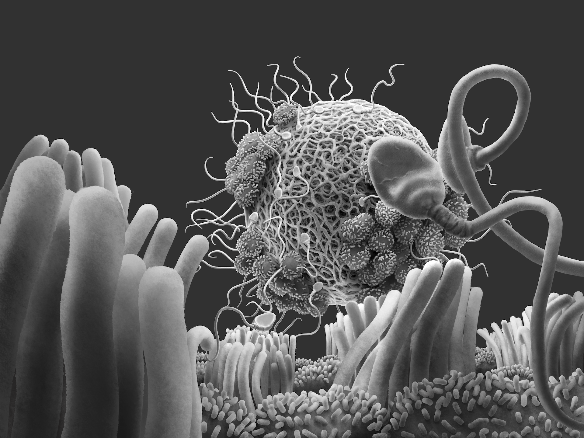
The appearance of the zona pellucida
The zona pellucida, well known as the covering of the egg for preventing polyspermy, i.e., preventing multiple sperm cells from entering the egg, has been usually perceived as a solid sheath enclosing the oocyte. This is partially true, because through light microscopy, the zona appears to be darkly coloured when stained, see figure 1.

Recent studies have shown that the zona appears to form a mesh like structure, and can have different types of appearances, i.e., spongy to solid in appearance (2). Although it may appear differently, questions are raised if they affect the outcome of fertilization in in-vitro studies (3). See figure 2.

The internal environment of the fallopian tube
The fallopian tube, also called the uterine tube, is the passage between the ovary to the uterus. It is here where the sperm cells usually cross paths with the egg complex and fertilization ensues. The tube is formed by two types of cells, termed as non-ciliated and ciliated cells. On looking closer, the non-ciliated cells exhibit numerous microvilli that may have several functions in cell signalling and interactions with the environment. See figure 3.

The cumulus cells and the oocyte
The cumulus cells are the granulosa cells of the corona radiata that covers the developing oocyte in the ovary. These cells have a lasting relationship with the oocyte. There appears an intimate structural relationship between the oocyte and the cumulus cells, where fine canalicular projections from the surface of the cumulus cells pierce the zona pellucida and form cell junctions with the microvilli of the oocyte (4). See figure 4.

The ECM secreted by the cumulus cells
Another fascinating fact is that the cumulus cells are also secretory in nature. It has been found that they secrete an extra cellular matrix (ECM), composed of several protein fibres and signalling molecules that allow the ciliated cells of the uterine tube to interact with the egg and possibly influence the direction of motility of the cilia (2).
Concluding remarks
This is a brief report on some findings that have been around. The illustration was made on procreate referencing current SEM studies on human oocytes and spermatozoa. The apperance via SEM studies shows a solid world due to powder coating the specimens with heavy metals to make it suitable for electron microscopy, and is one of the several visual models available for study. However, when viewed via other microscopic techniques, the zona will appear as a translucent structure.
It appears that by documenting cellular phenomena visually and spatially, newer models of reasoning can be developed to investigate the depths of such processes. Let us know what you think about these visual models and share your thoughts in the comments below.
References:
- Ekman AA, Chen JH, Guo J, McDermott G, Le Gros MA, Larabell CA. Mesoscale imaging with cryo‐light and X‐rays: Larger than molecular machines, smaller than a cell. Biology of the Cell. 2017 Jan;109(1):24-38.
- Familiari G., Relucenti M., Heyn R., Micara G., Correr S. (2006). Three-dimensional structure of the zona pellucida at ovulation. Microsc. Res. Tech. 69 415-426.
- Gupta SK. Human zona pellucida glycoproteins: binding characteristics with human spermatozoa and induction of acrosome reaction. Frontiers in cell and developmental biology. 2021 Feb 11;9:619868.
- Clarke HJ. Regulation of germ cell development by intercellular signaling in the mammalian ovarian follicle. Wiley Interdisciplinary Reviews: Developmental Biology. 2018 Jan;7(1):e294.
Liked something? leave your thoughts